


Page Under Maintenance
This page is currently under maintenance.
Please communicate at the hospital help desk for assistance.
Nuclear medicine is a specialized branch of diagnostic imaging that allows us to visualize organ function, detect early disease, and understand complex physiological processes inside the body. Unlike traditional radiology, which focuses on structural details, nuclear medicine provides a functional view of tissues and organs using a radioactive tracer and advanced tools like the gamma camera. This unique ability makes nuclear imaging a vital diagnostic tool for evaluating heart conditions, kidney disorders, thyroid diseases, bone problems, and various cancers. Through this comprehensive guide, we explore the principles, procedures, applications, and future innovations shaping the world of nuclear medicine.
Nuclear medicine technology allows us to observe how organs work in real time. By administering a radioactive tracer, we can track its movement and accumulation in targeted tissues, revealing underlying metabolic processes. This makes nuclear imaging incredibly powerful in diagnosing disorders before structural damage appears. The combination of radiopharmaceutical science and sensitive detectors makes this field one of the most precise diagnostic tools available today.
A radioactive tracer is a medically safe compound that behaves like natural molecules in the body. Once administered, it participates in biological processes and emits gamma rays that can be detected externally. These emissions create a map of organ function, showing areas of increased or decreased activity. Because the tracer binds to specific tissues, physicians can evaluate targeted organs with remarkable precision. This helps detect conditions long before they appear on CT or MRI scans, making the tracer an essential component of early diagnosis.
Diagnostic imaging in nuclear medicine focuses on monitoring the metabolic pathways of the tracer as it moves throughout the body. Regions that take up more tracer may indicate hyperactive tissue such as tumors or inflammation, while areas with insufficient uptake suggest reduced function or ischemia. This dynamic tracking allows clinicians to understand disease progression at a functional level. It also enables the identification of hidden abnormalities that traditional visual imaging might overlook, giving nuclear medicine a vital role in today’s diagnostics.
Functional changes occur before structural abnormalities, which means nuclear medicine can detect disease at its earliest stages. By assessing organ function rather than form, physicians can identify disorders such as kidney obstruction, thyroid imbalance, or cardiac ischemia long before symptoms escalate. Early functional diagnosis supports timely treatment and significantly improves outcomes. This predictive approach provides a deeper understanding of patient health and leads to personalized care strategies.
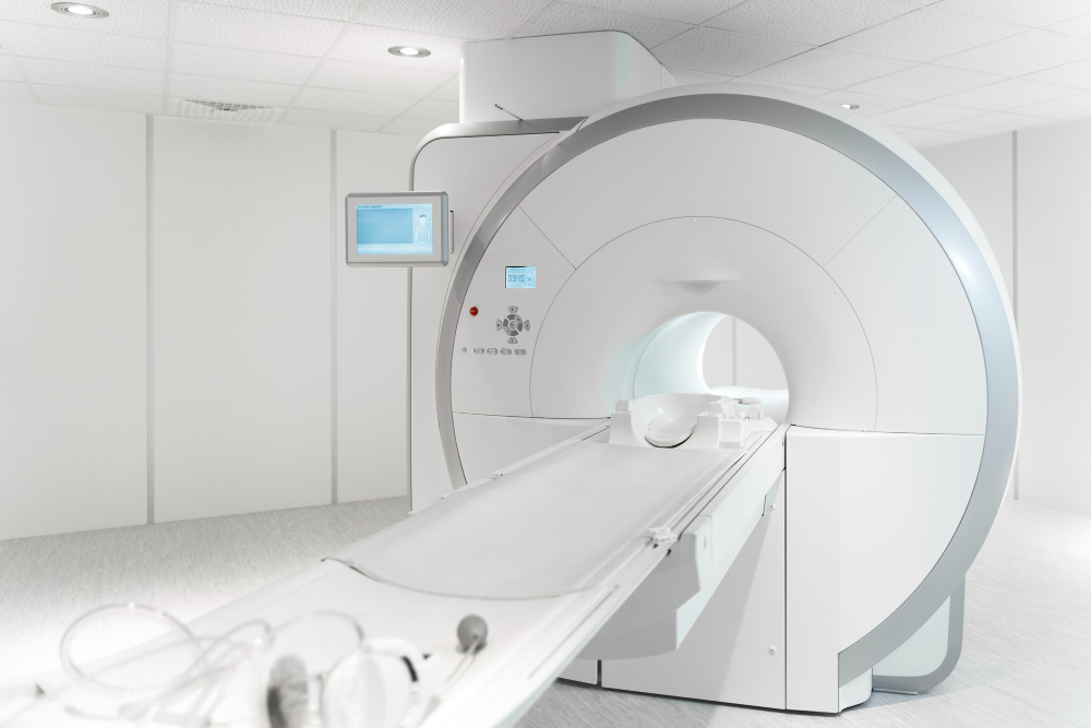
The gamma camera is central to nuclear medicine, converting gamma-ray emissions into diagnostic images. It uses advanced detectors to capture radiation safely and accurately. This technology transforms invisible energy into meaningful clinical data that helps physicians understand organ function in depth. The precision of the gamma camera ensures accurate evaluation of tracer distribution and enhances the quality of diagnostic results.
A gamma camera contains a scintillation crystal that reacts to gamma photons emitted from the tracer inside the body. When photons hit the crystal, flashes of light are produced and converted into electrical signals. These signals are then processed into images that reveal where the tracer accumulated. This process is fast, noninvasive, and painless, allowing physicians to evaluate functional patterns. The ability to record subtle changes in tracer absorption makes the gamma camera indispensable in modern nuclear imaging.
Modern gamma cameras incorporate multi-headed detectors that capture images from several angles simultaneously. This improves resolution, reduces scan time, and enhances diagnostic accuracy. Digital enhancements ensure that even low-level emissions are recorded clearly. With improved processing power and refined hardware, contemporary gamma cameras produce images that are detailed, reliable, and easier for physicians to interpret accurately.
Single Photon Emission Computed Tomography, or SPECT, uses the gamma camera in rotation to generate 3D images of internal organs. This advanced method shows depth, volume, and function with exceptional clarity. By reconstructing cross-sectional images, SPECT provides detailed insights into heart perfusion, brain function, and tumor behavior. It delivers a multidimensional view that supports thorough diagnosis and enhances clinical decision-making.
Nuclear medicine encompasses a wide range of tests, each designed to assess specific organs or conditions. These tests help diagnose diseases early, monitor treatment response, and guide clinical management. Because each tracer targets different tissues, the versatility of nuclear imaging makes it invaluable across many medical specialties.
A nuclear bone scan uses tracer uptake to reveal abnormalities in bone metabolism. Areas with high tracer absorption indicate increased metabolic activity caused by fractures, infections, arthritis, or tumors. Because functional changes appear before structural damage, bone scans identify disease earlier than X-rays. They are especially useful in detecting stress fractures, metastatic cancer, and unexplained bone pain that conventional imaging may miss.
Thyroid nuclear imaging evaluates how well the gland absorbs iodine-based tracers. This helps diagnose hyperthyroidism, hypothyroidism, goiters, and nodules. By comparing uptake patterns, physicians determine whether nodules are overactive or suspicious for cancer. The functional insights gained from these studies allow precise treatment planning, especially for patients undergoing thyroid therapy or follow-up assessments after intervention.
Renal scans evaluate kidney blood flow, filtration, and drainage using targeted radiopharmaceuticals. These tests identify obstructions, assess renal artery stenosis, and determine overall kidney function. By comparing the performance of each kidney individually, renal scans guide surgical planning and help monitor transplant health. They are essential in differentiating reversible blockages from irreversible damage, ensuring accurate treatment decisions.
Nuclear medicine plays a crucial role in evaluating the heart and other major organs. It reveals functional abnormalities that structural imaging often cannot detect. Through targeted tracers and precise imaging techniques, nuclear cardiology offers invaluable information on cardiac perfusion, tissue viability, and overall heart health.
Cardiac nuclear imaging assesses how well blood flows through the heart muscle during rest and stress. Tracer uptake patterns reveal whether certain regions receive inadequate blood supply, suggesting coronary artery disease. By comparing rest and stress results, physicians determine the severity of ischemia and plan appropriate interventions. This early detection can prevent heart attacks and long-term complications.
Nuclear imaging differentiates healthy tissue from scarred areas caused by previous heart attacks. This helps determine whether damaged regions still contain viable myocardium capable of recovering function. Identifying viable tissue is critical when considering procedures such as bypass surgery or angioplasty. These insights ensure that treatments are tailored for maximum benefit.
After treatment, nuclear imaging helps evaluate improvements in heart blood flow and muscle function. Changes in tracer distribution reflect the effectiveness of medical therapy, angioplasty, or surgical intervention. Regular monitoring provides a reliable way to track patient recovery and adjust therapeutic strategies accordingly.
A nuclear medicine procedure is simple, painless, and designed to provide accurate diagnostic information with minimal preparation. The process includes consultation, tracer administration, imaging, and post-scan care. Each step ensures safety and precision in capturing functional data.
Preparation varies depending on the test but may involve fasting, avoiding certain medications, or limiting caffeine. Proper preparation ensures that the radioactive tracer distributes correctly and produces reliable results. Patients are guided to stay hydrated and relaxed to maintain comfort. Clear communication with the medical team ensures a smooth and accurate imaging process.
Tracers are administered intravenously, orally, or through inhalation based on the organ being assessed. Once inside the body, the tracer circulates and accumulates in target tissues. Patients may wait minutes to hours depending on the study type. Because tracer doses are small and carefully controlled, the process is safe and well tolerated by most individuals.
During imaging, patients lie still while the gamma camera scans the targeted region. The camera detects gamma emissions and produces detailed functional images. The procedure is quiet, painless, and usually completed quickly. After the scan, physicians analyze the images to interpret tracer distribution and diagnose underlying conditions.
Nuclear medicine offers multiple advantages, including early disease detection, functional insights, and personalized diagnostics. Safety protocols ensure minimal radiation exposure, and ongoing advancements continue to improve imaging quality.
Because nuclear medicine detects physiological changes, it identifies disease earlier than structural imaging. This leads to faster interventions and improved patient outcomes. Functional imaging helps clarify complex disorders, making it indispensable in cardiac, renal, endocrine, and oncological evaluations. The precision of nuclear medicine supports more confident clinical decisions.
Radiation exposure from nuclear imaging is low and comparable to common diagnostic scans. The body naturally eliminates most radioactive tracer material within hours or days. Strict safety standards protect patients and ensure responsible tracer use. Nuclear medicine remains one of the safest functional imaging methods available today.
PET Scans Explained: How They Help Diagnose Diseases
Innovations such as PET-CT, PET-MRI, AI-driven diagnostics, and next-generation gamma camera are transforming nuclear medicine. These advancements enhance resolution, reduce scan time, and improve clinical accuracy. New radiopharmaceuticals are being developed to target diseases at the molecular level, paving the way for personalized medicine and therapeutics.
Scarless endoscopic mastectomy is a minimally invasive breast surgery technique performed through small incisions.
Yes, it is considered safe when performed by experienced surgeons who specialize in endoscopic procedures.
Recovery time usually ranges from two to four weeks, depending on the patient’s overall health and whether reconstruction is done simultaneously