


Page Under Maintenance
This page is currently under maintenance.
Please communicate at the hospital help desk for assistance.
A PET scan is one of the most advanced diagnostic imaging tools used in modern medicine. It helps doctors see how organs and tissues are functioning at a cellular level, giving insights far beyond what traditional imaging shows. PET scans help detect diseases early, monitor treatment response, and guide medical decisions. By understanding how these scans work, patients can feel more confident during their diagnostic journey.
A PET scan is a medical imaging test that captures metabolic activity inside the body using a radioactive tracer. The scan identifies how tissues absorb and use energy, which reveals early signs of disease before structural changes occur. Doctors rely on PET scans to diagnose cancer, evaluate heart disease, and study brain disorders. Because the scan focuses on cell behavior, it provides information that other tests may miss. PET technology is valuable because it helps detect abnormalities early, which can significantly improve treatment outcomes.
Positron emission tomography is a sophisticated imaging technique that tracks positrons emitted by a radiotracer. When these particles interact with electrons in the body, they produce signals captured by the scanner. This technology allows physicians to interpret metabolic patterns that help detect disease early and monitor progress. PET scans play a crucial role in oncology, neurology, and cardiology because they illustrate how well tissues are functioning rather than simply showing their shape. By revealing these functional changes, PET scans offer deeper, more actionable insights.
A standalone PET scan shows how organs function, but a PET/CT scan combines this information with detailed structural images from a CT scanner. This hybrid test helps doctors pinpoint exactly where abnormal metabolic activity occurs within the body’s anatomy. PET detects functional changes, while CT identifies precise physical locations. Together, they deliver highly accurate diagnostic information. A PET/CT scan is particularly useful in cancer staging, treatment planning, and monitoring therapy response because it combines two powerful imaging perspectives in one session.
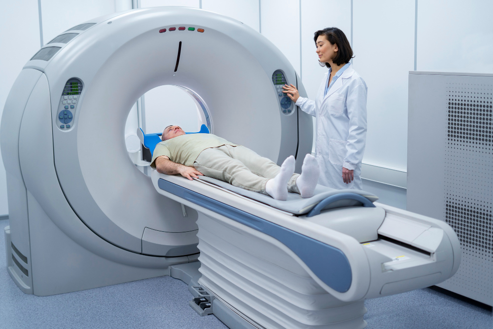
Understanding how a PET scan works can make the entire experience feel less intimidating. The process involves injecting a radiotracer, allowing it to travel through the body, and then using a scanner to detect emitted signals. These signals are converted into images that highlight metabolic activity.
A radiotracer is a small amount of radioactive material attached to a natural substance, such as glucose. When injected into the bloodstream, the tracer follows biological pathways and accumulates in tissues based on their metabolic activity. Highly active cells absorb more tracer, making them appear brighter in the images. This helps doctors identify abnormalities like tumors, inflammation, or damaged tissues.
The PET scan procedure begins with a radiotracer injection, followed by a resting period to allow the tracer to distribute throughout the body. During this time, patients are asked to relax and avoid movement, which ensures optimal image quality. The actual scanning process involves lying still on a table that slides into a circular scanner. The machine detects signals emitted by the tracer and forms them into images.
The PET scanner collects data from the radiotracer and converts it into detailed, color-coded images. Areas with high metabolic activity appear as bright regions, while low-activity areas look dimmer. Radiologists carefully analyze these patterns to determine whether they reflect normal or abnormal biological processes. Interpretation requires expertise because multiple conditions can cause similar imaging patterns.
A PET/CT scan is one of the most effective diagnostic tools because it provides a complete view of both function and structure. Doctors often rely on PET/CT results to diagnose cancer, evaluate heart disease, and investigate brain disorders. The scan’s ability to capture detailed metabolic information makes it one of the most accurate methods for disease detection.
Cancer cells consume energy at a much faster rate than normal cells, making them easy to detect with a PET scan. A PET/CT scan highlights these active areas, helping doctors locate tumors even when they are very small. The scan also shows if cancer has spread to lymph nodes or other organs. This information is essential for staging the disease and choosing the right treatment.
PET imaging plays a major role in diagnosing heart conditions because it evaluates blood flow and cell activity in the heart muscle. A PET scan can identify areas that are not receiving enough blood, helping detect coronary artery disease. It also shows whether damaged heart tissue is still alive and capable of recovery. This information is critical for planning treatments like angioplasty or bypass surgery.
Neurological diseases often change the brain’s metabolism long before structural abnormalities appear. PET scans are extremely helpful in diagnosing conditions such as Alzheimer’s disease, epilepsy, and Parkinson’s disease. In Alzheimer’s, for example, PET scans show reduced activity in specific brain regions. In epilepsy, PET imaging pinpoints the exact area where seizures originate.
Preparation is an important part of achieving accurate results in a PET scan. Because the tracer tracks metabolic activity, factors like eating, exercise, and medication can influence the exam. Understanding how to prepare helps ensure the scan goes smoothly and produces reliable images.
Before a PET scan, patients are usually asked to avoid strenuous exercise for 24 hours because physical exertion can increase metabolic activity. Fasting may also be required for several hours before the test, depending on the type of radiotracer used. At the imaging center, patients provide their medical history and medication list. Special precautions are taken for pregnant or breastfeeding women.
Diet plays a major role in PET scan accuracy. Patients are often advised to avoid sugary foods, caffeine, and carbohydrates the night before the scan. This helps stabilize blood sugar levels, which improves tracer distribution. For diabetic patients, instructions are adjusted to ensure medication and insulin levels remain balanced. During the appointment, patients are asked to remove jewelry, belts, and metal accessories.
The patient experience during a PET scan is straightforward and comfortable. After the radiotracer injection, the patient rests quietly for up to an hour while the tracer spreads through the body. When it is time for the scan, the patient lies on the table, which gently moves into the scanner. The machine silently collects data without causing discomfort. Patients must remain still during the scan to ensure clear images.
Although PET scans involve radiation, the levels used are low and carefully controlled. Medical professionals evaluate each patient to ensure the scan is safe and appropriate. Understanding the risks and safety precautions can help ease concerns.
The radiotracer used in PET scans is safe for most people. It contains a very small amount of radioactive material that decays quickly. The body naturally eliminates it within a few hours. Most patients experience no side effects, although some may briefly feel a cool sensation during injection. The tracer dose is carefully calculated to minimize exposure while still producing accurate images.
Radiation exposure during a PET scan is similar to that of a diagnostic CT scan. The total amount varies depending on the radiotracer and the area being scanned. Regulatory bodies set strict guidelines to ensure patient safety. Doctors only recommend PET imaging when the benefits clearly outweigh the risks. After the scan, patients naturally reduce radiation levels in their bodies as the tracer is eliminated. Staying hydrated can help speed up this process.
Although PET scans are safe for most people, certain individuals should avoid them unless medically necessary. Pregnant women are generally discouraged from undergoing PET scans due to potential risks to the fetus. Breastfeeding mothers may need to pause feeding temporarily. Patients with severe allergies or uncontrolled diabetes may require special preparation or alternative tests.
PET imaging is invaluable, but it also has limitations. Understanding both helps patients and healthcare providers make informed decisions about diagnosis and treatment.
PET imaging stands out because it detects disease early, often before physical symptoms appear. It provides detailed metabolic information that helps guide treatment decisions. PET scans also help doctors evaluate how well a treatment is working, allowing them to adjust therapies quickly. The technology reduces the need for invasive procedures in many cases.
Despite its strengths, PET imaging is not perfect. Certain conditions like inflammation or infection can create signals that resemble cancer activity. PET scans may not detect very small tumors or lesions that have low metabolic rates. Additionally, the cost of PET imaging may limit access for some patients. PET scanners require specialized facilities, meaning they are not always available in smaller hospitals.
PET scans are often compared with MRI, CT, and ultrasound. While MRI excels at capturing detailed pictures of soft tissue and CT provides clear structural images, PET scans reveal functional changes in tissues. This makes PET invaluable for detecting disease activity at an early stage. When combined with CT, PET scans create a complete picture that captures both structure and function.
Advancements in imaging technology continue to improve PET scans, making them faster, more accurate, and more patient-friendly. The future of PET imaging looks promising as researchers develop new techniques to enhance diagnostic capabilities.
Researchers are creating new radiotracers designed to target specific diseases more precisely. These tracer innovations allow doctors to study unique biological processes linked to cancer, infections, and neurological disorders. Personalized radiotracers tailored to a patient’s specific condition are also being explored. These advancements will likely lead to more accurate diagnoses and better treatment planning.
Modern PET scanners can capture images faster than previous generations. These advanced machines offer improved comfort, shorter scan times, and higher resolution. Some devices can scan the entire body in just minutes. Artificial intelligence is also improving the accuracy of PET image interpretation by identifying abnormal patterns earlier than the human eye.
PET imaging plays an essential role in precision medicine, which focuses on tailoring treatments to individual patients. PET scans reveal metabolic patterns that help predict how a patient will respond to therapy. This allows doctors to choose the most effective treatment while avoiding unnecessary procedures. As personalized medicine continues to evolve, PET imaging will become even more crucial in guiding individualized care.
PET scans, PET/CT scans , and Positron emission tomography have revolutionized the way diseases are diagnosed and monitored. Their ability to detect early metabolic changes provides doctors with unparalleled insights into organ function. From diagnosing cancer to evaluating heart and brain conditions, PET technology has become an essential tool for accurate and timely medical evaluation. While there are limitations, the benefits are extensive and continue to grow as technology advances.
No, PET scans are not painful. The only minor discomfort is the small injection for the radiotracer.
The radiotracer naturally leaves the body within a few hours, mostly through urine.
PET scans detect most cancers, but very small or slow-growing tumors may not always appear clearly.